


Diagnosis of prostatitis includes more than 5 mandatory and 4 additional procedures. Just a rectal exam of the prostate or an ultrasound cannot tell for sure if men have inflammation in the prostate. Because many urological diseases have a similar clinical picture and only a comprehensive differential study can rule out incorrect diagnoses.
How to pass an inspection
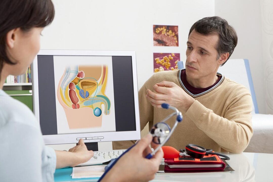
Men are recommended to have a preventive examination of the prostate by a urologist 1-2 times a year (prostatitis, adenoma and other pathologies of the prostate are asymptomatic in the first stages). If signs of illness appear, you should consult a specialist immediately. Such symptoms include pain in the lower abdomen and groin, difficulty urinating and erection.
The doctor begins by collecting complaints and anamnesis of the patient, and then conducts a general examination. The next step when prostatitis is suspected is a rectal exam (palpation of the prostate through a man's rectum). Finger research allows the doctor to evaluate the following parameters:
- The size of the prostate.
- surface (smooth or uneven).
- The density of the gland (soft or rocky).
- The presence or smoothness of the central groove.
- A man's sensitivity to probing the prostate (whether he feels pain).
Normally, the prostate should have easily palpable 2 symmetrical lobules and a central groove. The diameter of a healthy prostate is 2. 5-3. 5 cm, longitudinal - 2. 5-3 cm, the surface should be even, without pronounced tubercles, soft enough, but not loose. Any deviation from the listed characteristics means prostatitis, prostate adenoma, fibrosis, cancer or other pathologies of the genitourinary system.
analyses
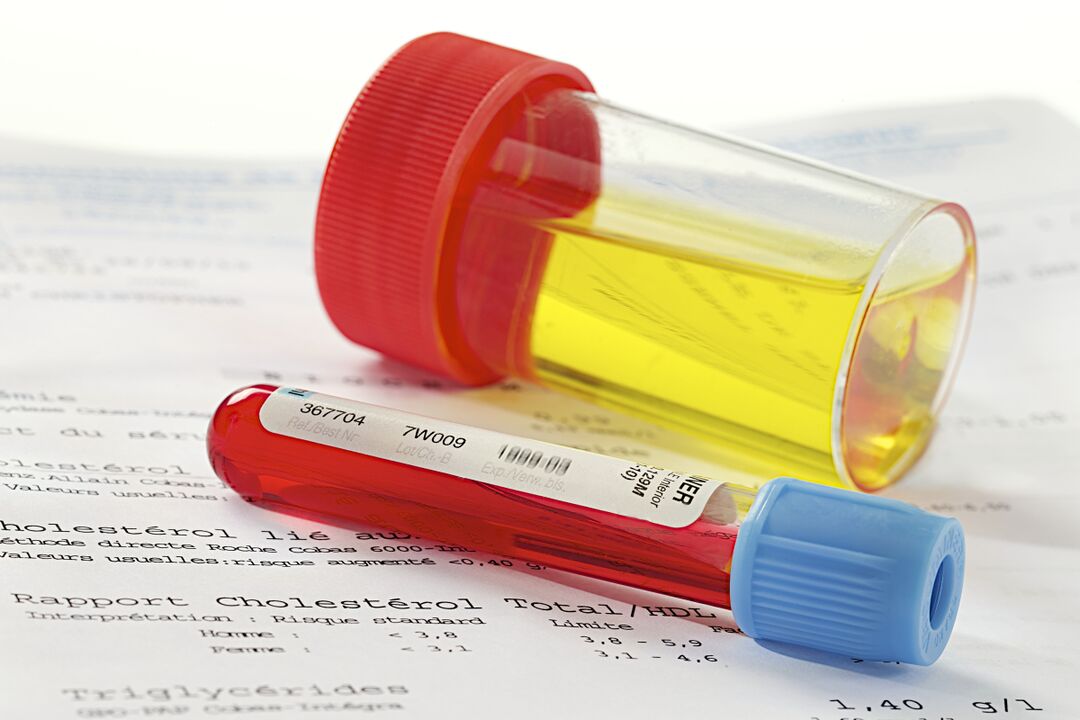
If rectal exam and history suggest prostatitis, the urologist's next move is to refer the patient for laboratory testing. According to clinical standards, the following types of examinations are mandatory:
- clinical urinalysis;
- general blood analysis;
- sowing urine for flora;
- when infection is detected, the sensitivity of pathogens to antibiotics is determined.
A complete blood count helps to confirm acute prostatitis - with this diagnosis, the number of neutrophils increases with a shift in the leukocyte formula to the left and a sharp decrease in the level of eosinophils. It is also possible to increase the ESR. Chronic inflammation is characterized by low hemoglobin levels (below 100 grams per liter of blood).
To rule out prostate cancer, a blood serum test for the level of PSA – a prostate-specific antigen – is carried out. Its increased amount indicates the presence of tumors, but does not determine their nature (benign or malignant). To find out this parameter, a biopsy of the prostate is performed with a histological study of the material obtained.
prostate secret
During a rectal examination of the prostate, the urologist pays attention to the secretion. Usually it is viscous, odorless and white in color. The maximum volume is 1-2 drops (3-5 ml). It should not contain any pus or blood impurities, as this is a sign of disease. The consistency of the juice matters - if it comes out in lumps, then the man has diverticulosis prostatitis. A more detailed examination of the material allows laboratory tests.
The microscopic and bacteriological study of the secret of the prostate is based on the counting of leukocytes, lecithin grains, amyloid bodies, macrophages, pathogenic and opportunistic organisms. Prostatitis is characterized by deviations:
- Acute prostatitis: the color of the secret is yellowish, the odor is sweetish, the pH is acidic, there are less than half of leukocytes and up to ¼ of epithelial cells.
- Chronic bacterial prostatitis: yellow or brown color, sour odor, acidic pH, less than half of leukocytes, macrophages (over 15), many amyloid bodies.
- Chronic nonbacterial prostatitis: the color is reddish, brown, there is no smell, leukocytes are normal, macrophages (10–20) are detected, there are many amyloid bodies.
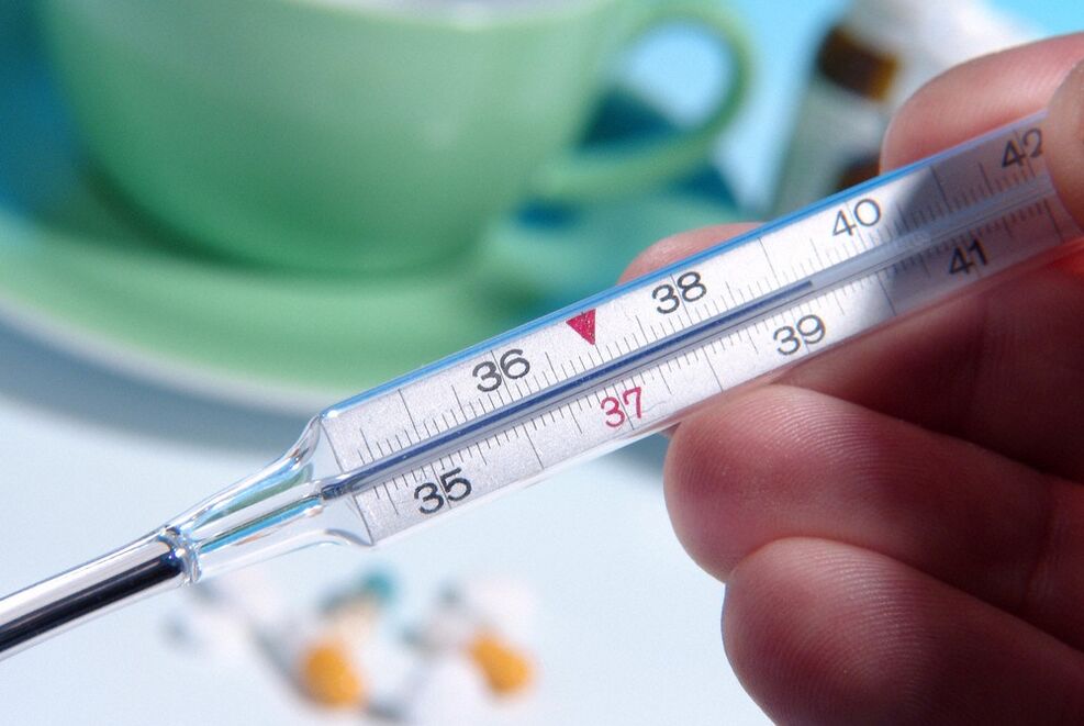
In some cases, studying the secret does not allow to detect prostatitis due to false indicators. Blurry data is displayed with inflammation in other organs and body temperature above 39 degrees. Taking material is not possible with contraindications to rectal massage (prostate juice is obtained by this method): with exacerbation of hemorrhoids, anal fissures, prostatic tuberculosis.
urine
The general and cytological analysis of urine does not require special preparation. A man needs to collect the material in a container in the morning before breakfast (it is better to buy a sterile plastic container at a pharmacy). A few hours before, the patient is not recommended to empty the bladder, and one should not take medicines and alcoholic beverages a day before.
With the catarrhal form of the disease, deviations from the norm in the general analysis of urine can not be observed. With prostatitis of the last stages, purulent threads are detected in the examined material, which fall out.

Examination of a man's urine allows for the diagnosis of leukocyturia (an increase in the level of leukocytes that occurs with inflammation). A urine culture is done to determine the type of pathogens. Signs of pathogens in the urine appear with infectious prostatitis or complications such as cystitis, urethritis or pelvic inflammatory disease.
Swabs from the urethra
A smear from the urethra is a type of examination that confirms inflammation caused by pathogens such as trichomonads, gonococci, candida. It is prescribed when chronic pelvic pain syndrome, itching in the groin, rash on the penis, difficulty in urinating are observed. Examination of the material taken allows differential diagnosis - to distinguish between prostatitis, urethritis or sexually transmitted diseases, which often have similar symptoms or occur simultaneously.
The disease can only be diagnosed with a correctly taken smear test. A man must abstain from sex for 2 days before ingesting the material. Do not go to the toilet an hour before the procedure. If the patient is taking NSAIDs or antibiotics, then there is no point in doing this analysis - the data will be wrong.
spermogram
Spermogram - analysis of a man's ejaculate. In addition to prostatitis, diseases of the seminal vesicles, testicles and infertility are also diagnosed in this way. The material submitted by a man with a body temperature not higher than 39 degrees, who does not take antibiotics and abstains from sexual intercourse for 2-3 days, is correct. A prostate massage is not recommended the day before sperm donation.
The spermogram includes three types of studies. Macroscopic analysis involves examining the volume, color, viscosity and liquefaction time of the seed. The microscopic examination shows the quantity and quality of the sperm. Biochemical analysis determines the concentration in the ejaculate of fructose, zinc, alpha-glucosidase, L-carnitine. Antisperm antibodies can be detected in bacterial prostatitis.
With prostatitis, a spermiogram can reveal a number of abnormalities. For example, a reduced volume of semen (less than 1. 5 ml), a low concentration of spermatozoa in 1 ml (less than 15 million), asthenozoospermia (more than 40% of immobile sperm), akinospermia (more than 32% of immobile sperm ).
prostate tissue
When examining an enlarged prostate, it is not always possible to understand the nature of the seals and extensions using a rectal exam, urine and blood tests. It can be a benign pathology (adenoma, prostatitis) or malignant (cancer). Accurate diagnosis helps microscopic examination of prostate tissues obtained by biopsy.
The procedure is carried out as follows: the patient is introduced transrectally with an ultrasonic device sensor, at the end of which there is a gun with a biopsy needle. A microscopic section of glandular tissue is cut off with a sharp point and sent to the laboratory for examination. The study is carried out using the method of comparing the parameters of the material with the norms from the Gleason table.
With congestive, viral or bacterial prostatitis, the glandular cells look reduced, the amount of connective tissue in the intercellular space is increased. Atypical cells with altered nuclei are not observed. When a man has prostate cancer, the glandular cells grow large and gather in clusters, their abnormal modifications are revealed.
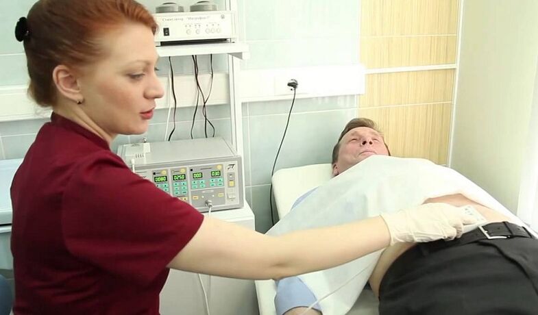
Ultrasound, MRI and other methods
Instrumental studies are carried out to confirm the diagnosis, as well as to determine the stage of development and features of the course of the disease. With pathologies of the pelvic organs, the following methods of examination are used:
- traditional ultrasound;
- transrectal ultrasound;
- magnetic resonance imaging (MRI);
- CT scan.
These methods allow you to find out the shape, thickness, width, length of the prostate, its mass, structural uniformity, echogenicity, vascularization (vascular pattern). These parameters are needed to determine urological pathologies: ultrasound, CT and MRI reveal inflammatory, proliferative, oncological diseases of the prostate.
Classic ultrasound has the greatest inaccuracy, but this method continues to be used because it is easy to use and affordable. Transrectal ultrasound is considered the "gold standard" for detecting prostatitis, but prostate cancer is difficult to detect this way (especially in its early stages). MRI and CT have the highest accuracy in identifying tumors, but these are complex and expensive procedures, therefore they are carried out when other research methods show a high probability of oncology.
examination at home
The prostate can be examined at home and the primary symptoms of urological pathologies can be identified. Of course, this is not a diagnosis of chronic prostatitis, since the cause of the enlarged gland cannot be reliably determined. But the presence of alarming signs with an independent examination of one's own body is an important reason to immediately contact a urologist.
Just like that, without the need to conduct self-diagnosis, it's not worth it. Indications to be examined at home are:
- Impaired urodynamics (frequent urge to urinate).
- Weak stream, inability to completely empty the bladder.
- Abdominal or groin discomfort (e. g. painful urination).
- Decreased sexual desire, weakening of the erection.
- Purulent impurities or discoloration of the urine to white, brown.
- spermatorrhea or prostorrhea (discharge from the penis).
At home, the examination is carried out according to the same scheme as in the doctor's office. First, a man needs to clean the intestines - in 10-12 hours do an enema or take laxatives. Take a bath immediately before the procedure. Then lie on your side, bend your knees, insert your index finger into the rectum (previously put a fingertip and smear it with petroleum jelly).
A digital rectal exam is performed by probing the posterior wall of the colon and capturing the adjacent prostate. The gland is easy to spot - it feels like a small walnut. Bad symptoms: enlarged prostate, non-circular shape, presence of tubercles, pain on probing.These signs signal inflammation or some other pathological process in the prostate. If they are identified, be sure to go to the urologist, as a more accurate diagnosis and treatment plan is required.